

Can You Repair A St Jude Aortic Valve
| Artificial heart valve | |
|---|---|
 Different types of bogus center valves[one] | |
| Specialty | cardiology |
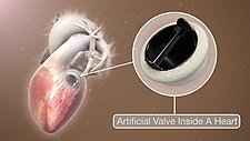
An bogus heart valve is a one-way valve implanted into a person'south heart to supplant a eye valve that is non functioning properly (valvular heart disease). Artificial middle valves can be separated into three broad classes: mechanical heart valves, bioprosthetic tissue valves and engineered tissue valves.
The human eye contains four valves: tricuspid valve, pulmonary valve, mitral valve and aortic valve. Their main purpose is to keep blood flowing in the proper direction through the eye, and from the heart into the major blood vessels connected to it (the pulmonary artery and the aorta). Center valves can malfunction for a variety of reasons, which tin impede the menstruum of blood through the valve (stenosis) and/or let blood flow backwards through the valve (regurgitation). Both processes put strain on the eye and may lead to serious problems, including heart failure. While some dysfunctional valves can exist treated with drugs or repaired, others demand to be replaced with an bogus valve.[2]
Background [edit]
3D Medical Blitheness still shot of Bogus Heart Valve
A heart contains four valves (tricuspid, pulmonary, mitral and aortic valves) which open up and close as blood passes through the heart.[3] Claret enters the heart in the correct atrium and passes through the tricuspid valve to the right ventricle. From there, claret is pumped through the pulmonary valve to enter the lungs. After being oxygenated, blood passes to the left atrium, where is it pumped through the mitral valve to the left ventricle. The left ventricle pumps blood to the aorta through the aortic valve.
In that location are many potential causes of heart valve damage, such equally birth defects, age related changes, and furnishings from other disorders, such as rheumatic fever and infections causing endocarditis. High blood pressure and eye failure which can enlarge the heart and arteries, and scar tissue tin can form after a heart assail or injury.[4]
The three main types of artificial heart valves are mechanical, biological (bioprosthetic/tissue), and tissue-engineered valves. In the US, UK and the European union, the near common type of artificial heart valve is the bioprosthetic valve. Mechanical valves are more than commonly used in Asia and Latin America.[ commendation needed ] Companies that manufacture center valves include Edwards Lifesciences,[five] Medtronic,[6] Abbott (St. Jude Medical),[vii] LivaNova,[eight] CryoLife,[nine] and LifeNet Health.[10]
Mechanical valves [edit]
Mechanical valves come in three principal types – caged ball, tilting-disc and bileaflet – with various modifications on these designs.[11] Caged brawl valves are no longer implanted.[12] Bileaflet valves are the most common type of mechanical valve implanted in patients today.[13]
Caged ball valves [edit]

The kickoff bogus middle valve was the caged ball valve, a type of ball check valve, in which a ball is housed inside a cage. When the heart contracts and the blood force per unit area in the bedchamber of the eye exceeds the pressure on the outside of the chamber, the ball is pushed against the cage and allows blood to flow. When the middle finishes contracting, the pressure inside the bedroom drops and the ball moves back against the base of the valve forming a seal.
In 1952, Charles A. Hufnagel implanted caged ball heart valves into ten patients (six of whom survived the performance), marking the first success in prosthetic centre valves.[ commendation needed ] A similar valve was invented by Miles 'Lowell' Edwards and Albert Starr in 1960, unremarkably referred to as the Starr-Edwards silastic ball valve.[14] This consisted of a silicone ball enclosed in a methyl metacrylate cage welded to a band. The Starr-Edwards valve was showtime implanted in a human on Baronial 25, 1960, and was discontinued by Edwards Lifesciences in 2007.[14]
Caged ball valves are strongly associated with blood clot formation, so people who have one required a loftier degree of anticoagulation, unremarkably with a target INR of three.0–4.5.[15]
Tilting-disc valves [edit]

Introduced in 1969, the first clinically bachelor tilting-disc valve was the Bjork-Shiley valve.[16] Tilting‑disc valves, a blazon of swing check valve, are made of a metallic ring covered by an ePTFE textile. The metal ring holds, by means of two metallic supports, a disc that opens when the heart beats to let claret flow through, then closes again to preclude blood flowing backwards. The disc is usually made of an extremely difficult carbon textile (pyrolytic carbon), enabling the valve to function for years without wearing out.[ citation needed ]
Bileaflet valves [edit]
Introduced in 1979, bileaflet valves are made of two semicircular leaflets that circumduct around struts attached to the valve housing. With a larger opening than caged brawl or tilting-disc valves, they carry a lower take a chance of blood clots. They are, withal, vulnerable to blood backflow.[ citation needed ]
Advantages of mechanical valves [edit]
The major reward of mechanical valves over bioprosthetic valves is their greater durability.[17] Made from metal and/or pyrolytic carbon,[11] they tin can last 20–30 years.[17]
Disadvantages of mechanical valves [edit]
One of the major drawbacks of mechanical heart valves is that they are associated with an increased risk of blood clots. Clots formed past red claret cell and platelet damage tin can cake blood vessels leading to stroke. People with mechanical valves need to take anticoagulants (claret thinners), such as warfarin, for the residuum of their life.[17] Mechanical centre valves can also crusade mechanical hemolytic anemia, a condition where the cherry blood cells are damaged as they pass through the valve.[ citation needed ] Cavitation, the rapid formation of microbubbles in a fluid such as blood due to a localized drib of pressure, can lead to mechanical middle valve failure,[eighteen] so cavitation testing is an essential office of the valve design verification procedure.
Many of the complications associated with mechanical middle valves can be explained through fluid mechanics. For example, claret clot germination is a side outcome of high shear stresses created by the design of the valves. From an technology perspective, an platonic heart valve would produce minimal pressure drops, have pocket-sized regurgitation volumes, minimize turbulence, reduce prevalence of high stresses, and not create catamenia separations in the vicinity of the valve.[ citation needed ]
Implanted mechanical valves can crusade foreign body rejection. The claret may coagulate and eventually result in a hemostasis. The usage of anticoagulation drugs will exist interminable to preclude thrombosis.[19] [ non-chief source needed ]
Bioprosthetic tissue valves [edit]
Bioprosthetic valves are usually made from animal tissue (heterograft/xenograft) attached to a metal or polymer support.[12] Bovine (moo-cow) tissue is most commonly used, just some are fabricated from porcine (pig) tissue.[20] [ non-primary source needed ] The tissue is treated to forbid rejection and calcification.[ citation needed ]
Alternatives to animal tissue valves are sometimes used, where valves are used from human donors, as in aortic homografts and pulmonary autografts. An aortic homograft is an aortic valve from a man donor, retrieved either after their decease or from a heart that is removed to be replaced during a centre transplant.[13] A pulmonary autograft, also known as the Ross procedure, is where the aortic valve is removed and replaced with the patient'south own pulmonary valve (the valve between the correct ventricle and the pulmonary artery). A pulmonary homograft (a pulmonary valve taken from a cadaver) is then used to replace the patient'south ain pulmonary valve. This process was first performed in 1967 and is used primarily in children, equally information technology allows the patient'due south own pulmonary valve (at present in the aortic position) to grow with the child.[xiii]
Advantages of bioprosthetic heart valves [edit]
Bioprosthetic valves are less likely than mechanical valves to cause blood clots, so do not require lifelong anticoagulation. As a result, people with bioprosthetic valves have a lower take chances of bleeding that those with mechanical valves.[17]
Disadvantages of bioprosthetic heart valves [edit]
Tissue valves are less durable than mechanical valves, typically lasting ten–20 years.[21] This means that people with bioprosthetic valves have a college incidence of requiring another aortic valve replacement in their lifetime.[17] Bioprosthetic valves tend to deteriorate more quickly in younger patients.[22]
In recent years, scientists have developed a new tissue preservation applied science, with the aim of improving the durability of bioprosthetic valves. In sheep and rabbit studies, tissue preserved using this new technology had less calcification than command tissue.[23] A valve containing this tissue is at present marketed, but long-term durability data in patients are not all the same available.[24] [ non-primary source needed ]
Current bioprosthetic valves lack longevity, and will calcify over time.[25] When a valve calcifies, the valve cusps become stiff and thick and cannot close completely.[25] Moreover, bioprosthetic valves can't abound with or adjust to the patient: if a child has bioprosthetic valves they will need to get the valves replaced several times to fit their concrete growth.[25]
Tissue-engineered valves [edit]
For over 30 years researchers have been trying to grow heart valves in vitro.[26] These tissue‑engineered valves involve seeding man cells on to a scaffold.[26] The two main types of scaffold are natural scaffolds, such every bit decellularized tissue, or scaffolds made from degradable polymers.[27] The scaffold acts as an extracellular matrix, guiding tissue growth into the correct 3D structure of the heart valve.[27] [26] Some tissue-engineered eye valves accept been tested in clinical trials,[27] simply none are commercially available.
Tissue engineered heart valves tin exist person-specific and 3D modeled to fit an individual recipient[28] 3D printing is used because of its loftier accurateness and precision of dealing with different biomaterials.[28] Cells that are used for tissue engineered centre valves are expected to secrete the extracellular matrix (ECM).[25] Extracellular matrix provides back up to maintain the shape of the valves and determines the cell activities.[29]
Scientists can follow the construction of eye valves to produce something that looks like to them, but since tissue engineered valves lack the natural cellular basis, they either fail to perform their functions like natural eye valves, or function when they are implanted but gradually dethrone over fourth dimension.[ citation needed ] An platonic tissue engineered heart valve would be non‐thrombogenic, biocompatible, durable, resistant to calcification, grow with the surrounding middle, and exhibit a physiological hemodynamic profile.[30] To achieve these goals, the scaffold should exist carefully called—in that location are three main candidates: decellularized ECM (xenografts or homografts), natural polymers, and synthetic polymers.[30]
Differences between mechanical and tissue valves [edit]
Mechanical and tissue valves are made of different materials. Mechanical valves are generally made of titanium and carbon.[31] Tissue valves are made upward of human being or fauna tissue. The valves equanimous of human tissue, known as allografts or homografts, are from donors' homo hearts.[31]
Mechanical valves tin can be a improve selection for younger people and people at risk of valve deterioration due to its durability. It is as well preferable for people who are already taking blood thinners and people who would be unlikely to tolerate another valve replacement performance.[ citation needed ]
Tissue valves are better for older historic period groups equally another valve replacement operation may non be needed in their lifetime. Due to the risk of forming blood clots for mechanical valves and severe bleeding as a major side effect of taking claret-thinning medications, people who have a risk of blood bleeding and are not willing to take warfarin may also consider tissue valves. Other patients who may be more than suitable for tissue valves are people who have other planned surgeries and unable to take blood-thinning medications. People who plan to go significant may also consider tissue valves as warfarin causes risks in pregnancy.[ citation needed ]
Functional requirements of artificial centre valves [edit]
An artificial heart valve should ideally function like a natural eye valve.[12] The performance of natural centre valves is characterized by many advantages:
- Minimal regurgitation – This means that the amount of blood leaking backwards through the valve as information technology closes is small. Some degree of valvular regurgitation is inevitable and natural, up to around 5 ml per crush.[32] Nevertheless, several center valve pathologies (eastward.1000. rheumatic endocarditis) may lead to clinically significant valvular regurgitation. A desirable characteristic of heart valve prostheses is that regurgitation is minimal over the full range of physiological eye function.
- Minimal transvalvular pressure gradient – Whenever a fluid flows through a restriction, such every bit a valve, a force per unit area gradient arises over the restriction. This force per unit area gradient is a result of the increased resistance to flow through the restriction. Natural heart valves have a low transvalvular force per unit area slope every bit they nowadays little obstruction to the period through themselves, commonly less than sixteen mmHg. A desirable characteristic of heart valve prostheses is that their transvalvular pressure gradient is as modest as possible.
- Not-thrombogenic – Natural heart valves are lined with an endothelium comparable with the endothelium lining the heart chambers, and so they are not normally thrombogenic (i.due east. they don't crusade claret clots). Blood clots tin be hazardous because they can social club in, and cake, downstream arteries (e.g. coronary arteries, leading to heart assault [myocardial infarction]; or cerebral arteries, leading to stroke). A desirable characteristic of artificial heart valves is that they are non- or minimally thrombogenic.
- Self-repairing – Valve leaflets retain some capacity for repair thanks to regenerative cells (e.g. fibroblasts) in the connective tissue from which the leaflets are composed. As the human being centre beats approximately three.four×ten9 times during a typical human lifespan, this limited only nevertheless present repair chapters is critically important. No heart valve prostheses tin can currently cocky-repair, only tissue-engineered valves may eventually offer such capabilities.[27]
Bogus heart valve repair [edit]
Artificial heart valves are expected to last from x to thirty years.[17]
The well-nigh common problems with artificial heart valves are various forms of degeneration, including gross billowing of leaflets, ischemic mitral valve pathology, and minor chordal lengthening.[25] The repairing process of the artificial heart valve regurgitation and stenosis usually requires an open-center surgery, and a repair or partial replacement of regurgitant valves is normally preferred.[25]
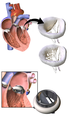
Researchers are investigating catheter-based surgery that allows repair of an bogus eye valve without large incisions.[33]
Additional images [edit]
-
3D Rendering of Mechanical Valve
-

3D Rendering of Mechanical Valve (St. Francis model)
Encounter also [edit]
- Artificial eye
- Mitral valve replacement
- Aortic valve replacement
References [edit]
- ^ Kostrzewa B, Rybak Z (2013). "[History, present and future of biomaterials used for artificial eye valves]". Polimery W Medycynie. 43 (three): 183–9. PMID 24377185.
- ^ Baumgartner H, Falk Five, Bax JJ, De Bonis Chiliad, Hamm C, Holm PJ, et al. (September 2022). "2017 ESC/EACTS Guidelines for the direction of valvular heart disease". European Middle Periodical. 38 (36): 2739–2791. doi:ten.1093/eurheartj/ehx391. PMID 28886619.
- ^ "Eye Valve Replacement: Which Type Is Best for You?". Health Essentials from Cleveland Clinic. 2022-06-14. Retrieved 2020-08-04 .
- ^ Muraru D, Anwar AM, Song J (Dec 2022). "Eye valve disease: tricuspid valve disease". Oxford Medicine Online. doi:10.1093/med/9780198726012.003.0037.
- ^ "Surgical aortic middle valves | Edwards Lifesciences". world wide web.edwards.com . Retrieved 2019-07-29 .
- ^ Medtronic. "Middle Valve Therapies - Surgical Replacement". www.medtronic.com . Retrieved 2019-07-29 .
- ^ "Surgical Valves | Trifecta GT Valve and Epic Mitral Valve". www.cardiovascular.abbott . Retrieved 2019-07-29 .
- ^ "LivaNova". world wide web.livanova.com . Retrieved 2019-07-29 . [ permanent expressionless link ]
- ^ "On-X Heart Valves". CryoLife, Inc . Retrieved 2019-07-29 .
- ^ "Cardiac | LifeNet Health". world wide web.lifenethealth.org . Retrieved 2019-07-29 .
- ^ a b Gott VL, Alejo DE, Cameron DE (December 2003). "Mechanical heart valves: 50 years of development". The Register of Thoracic Surgery. 76 (six): S2230-9. doi:10.1016/j.athoracsur.2003.09.002. PMID 14667692.
- ^ a b c Pibarot P, Dumesnil JG (Feb 2009). "Prosthetic heart valves: selection of the optimal prosthesis and long-term management". Circulation. 119 (7): 1034–48. doi:10.1161/CIRCULATIONAHA.108.778886. PMID 19237674.
- ^ a b c Bloomfield P (June 2002). "Choice of centre valve prosthesis". Heart. 87 (vi): 583–nine. doi:10.1136/heart.87.vi.583. PMC1767148. PMID 12010950.
- ^ a b Matthews AM (1998). "The development of the Starr-Edwards eye valve". Texas Center Institute Journal. 25 (4): 282–93. PMC325574. PMID 9885105.
- ^ Goldsmith I, Turpie AG, Lip GY (November 2002). "Valvar heart illness and prosthetic heart valves". BMJ. 325 (7374): 1228–31. doi:10.1136/bmj.325.7374.1228. PMC1124694. PMID 12446543.
- ^ Sunday JC, Davidson MJ, Lamy A, Eikelboom JW (August 2009). "Antithrombotic management of patients with prosthetic heart valves: current prove and future trends". Lancet. 374 (9689): 565–76. doi:ten.1016/S0140-6736(09)60780-7. PMID 19683642. S2CID 43661491.
- ^ a b c d eastward f Tillquist MN, Maddox TM (Feb 2022). "Cardiac crossroads: deciding between mechanical or bioprosthetic middle valve replacement". Patient Preference and Adherence. 5: 91–9. doi:10.2147/PPA.S16420. PMC3063655. PMID 21448466.
- ^ Johansen P (September 2004). "Mechanical heart valve cavitation". Proficient Review of Medical Devices. ane (one): 95–104. doi:10.1586/17434440.1.1.95. PMID 16293013. S2CID 27933945.
- ^ Namdari G, Eatemadi A (December 2022). "Nanofibrous bioengineered heart valve-Application in paediatric medicine". Biomedicine & Pharmacotherapy. 84: 1179–1188. doi:ten.1016/j.biopha.2016.x.058. PMID 27780149.
- ^ Hickey GL, Grant SW, Bridgewater B, Kendall S, Bryan AJ, Kuo J, Dunning J (June 2022). "A comparison of outcomes between bovine pericardial and porcine valves in 38,040 patients in England and Wales over 10 years". European Periodical of Cardio-Thoracic Surgery. 47 (half-dozen): 1067–74. doi:x.1093/ejcts/ezu307. PMID 25189704.
- ^ Harris C, Croce B, Cao C (July 2022). "Tissue and mechanical heart valves". Annals of Cardiothoracic Surgery. 4 (4): 399. doi:10.3978/6884 (inactive 28 February 2022). PMC4526499. PMID 26309855.
{{cite journal}}: CS1 maint: DOI inactive as of February 2022 (link) - ^ Johnston DR, Soltesz EG, Vakil North, Rajeswaran J, Roselli EE, Sabik JF, et al. (April 2022). "Long-term immovability of bioprosthetic aortic valves: implications from 12,569 implants". The Register of Thoracic Surgery. 99 (4): 1239–47. doi:x.1016/j.athoracsur.2014.x.070. PMC5132179. PMID 25662439.
- ^ Flameng W, Hermans H, Verbeken E, Meuris B (January 2022). "A randomized assessment of an avant-garde tissue preservation technology in the juvenile sheep model". The Journal of Thoracic and Cardiovascular Surgery. 149 (i): 340–5. doi:ten.1016/j.jtcvs.2014.09.062. PMID 25439467.
- ^ Bartuś One thousand, Litwinowicz R, Kuśmierczyk M, Bilewska A, Bochenek Thou, Stąpór M, et al. (2017-12-19). "Primary safety and effectiveness feasibility study subsequently surgical aortic valve replacement with a new generation bioprosthesis: 1-year outcomes". Kardiologia Polska. 76 (iii): 618–624. doi:10.5603/KP.a2017.0262. PMID 29297188. S2CID 4061454.
- ^ a b c d e f Hasan A, Saliba J, Pezeshgi Modarres H, Bakhaty A, Nasajpour A, Mofrad MR, Sanati-Nezhad A (October 2022). "Micro and nanotechnologies in heart valve tissue engineering". Biomaterials. 103: 278–292. doi:10.1016/j.biomaterials.2016.07.001. PMID 27414719.
- ^ a b c Stassen OM, Muylaert DE, Bouten CV, Hjortnaes J (September 2022). "Current Challenges in Translating Tissue-Engineered Heart Valves". Current Handling Options in Cardiovascular Medicine. 19 (nine): 71. doi:10.1007/s11936-017-0566-y. PMC5545463. PMID 28782083.
- ^ a b c d Blum KM, Drews JD, Breuer CK (June 2022). "Tissue-Engineered Center Valves: A Call for Mechanistic Studies". Tissue Engineering. Part B, Reviews. 24 (3): 240–253. doi:10.1089/ten.teb.2017.0425. PMC5994154. PMID 29327671.
- ^ a b Theus Every bit, Tomov ML, Cetnar A, Lima B, Nish J, McCoy 1000, Mahmoudi G, Serpooshan Five (2019-06-01). "Biomaterial approaches for cardiovascular tissue engineering". Emergent Materials. ii (ii): 193–207. doi:x.1007/s42247-019-00039-iii. ISSN 2522-574X. S2CID 201165755.
- ^ Hay ED (2013-11-11). Cell Biological science of Extracellular Matrix (Second ed.). Springer Science & Concern Media. ISBN978-ane-4615-3770-0.
- ^ a b Nachlas AL, Li S, Davis ME (Dec 2022). "Developing a Clinically Relevant Tissue Engineered Heart Valve-A Review of Electric current Approaches". Advanced Healthcare Materials. six (24): 1700918. doi:ten.1002/adhm.201700918. PMID 29171921. S2CID 2339263.
- ^ a b Schmidt JB, Tranquillo RT (2013). Tissue-Engineered Heart Valves. Eye Valves. Boston, MA: Springer U.s.a.. pp. 261–280. doi:10.1007/978-1-4614-6144-9_11. ISBN978-one-4614-6143-2.
- ^ Kasegawa H, Iwasaki K, Kusunose Southward, Tatusta R, Doi T, Yasuda H, Umezu M (January 2022). "Assessment of a novel stentless mitral valve using a pulsatile mitral valve simulator". The Journal of Eye Valve Illness. 21 (1): 71–5. PMID 22474745.
- ^ Bezuidenhout D, Williams DF, Zilla P (January 2022). "Polymeric centre valves for surgical implantation, catheter-based technologies and heart assist devices". Biomaterials. 36: six–25. doi:10.1016/j.biomaterials.2014.09.013. PMID 25443788.
Farther reading [edit]
- Bendet I, Morozov SM, Skumin VA (June 1980). "[Psychological aspects of the rehabilitation of patients subsequently the surgical treatment of heart defects]" Психологические аспекты реабилитации больных после хирургического лечения пороков сердца [Psychological aspects of the rehabilitation of patients subsequently the surgical treatment of heart defects]. Kardiologiia (in Russian). 20 (6): 45–51. PMID 7392405.
- Skumin VA (September 1979). "[Nurse'due south office in doc-psychological rehabilitation of patients with artificial middle valves]". Meditsinskaia Sestra. 38 (9): 44–5. PMID 259874.
- Skumin VA (1982). "[Nonpsychotic mental disorders in patients with caused middle defects earlier and after surgery (review)]". Zhurnal Nevropatologii I Psikhiatrii Imeni Southward.S. Korsakova. 82 (eleven): 130–v. PMID 6758444.
- Klepetko Westward, Moritz A, Mlczoch J, Schurawitzki H, Domanig Eastward, Wolner E (January 1989). "Leaflet fracture in Edwards-Duromedics bileaflet valves". The Journal of Thoracic and Cardiovascular Surgery. 97 (1): 90–4. doi:ten.1016/S0022-5223(19)35130-X. PMID 2911200.
- Podesser BK, Khuenl-Brady G, Eigenbauer E, Roedler S, Schmiedberger A, Wolner E, Moritz A (May 1998). "Long-term results of heart valve replacement with the Edwards Duromedics bileaflet prosthesis: a prospective ten-year clinical follow-upwards". The Journal of Thoracic and Cardiovascular Surgery. 115 (five): 1121–9. doi:10.1016/s0022-5223(98)70412-x. PMID 9605082.
- Knapp RJ, Daily JW, Hammitt FG (1970). Cavitation. New York: McGraw-Loma Int. Book Co.
- Lim WL, Chew YT, Depression HT, Foo WL (September 2003). "Cavitation phenomena in mechanical eye valves: the role of squeeze catamenia velocity and contact area on cavitation initiation betwixt two impinging rods". Journal of Biomechanics. 36 (9): 1269–lxxx. doi:x.1016/s0021-9290(03)00161-1. PMID 12893035.
- Bluestein D, Einav Due south, Hwang NH (November 1994). "A squeeze menstruum phenomenon at the closing of a bileaflet mechanical eye valve prosthesis". Periodical of Biomechanics. 27 (eleven): 1369–78. doi:10.1016/0021-9290(94)90046-ix. PMID 7798287.
- Graf T, Fischer H, Reul H, Rau G (March 1991). "Cavitation potential of mechanical heart valve prostheses". The International Periodical of Artificial Organs. 14 (3): 169–74. doi:x.1177/039139889101400309. PMID 2045192. S2CID 23086590.
- Kafesjian R, Wieting DW, Ely J, Chahine GL, Frederick GS, Watson RE (1990). "Characterization of Cavitation Potential of Pyrolitic Carbon". In Bodnar E (ed.). Surgery for Heart Valve Illness: Proceedings of the 1989 Symposium. ICR. pp. 509–16. ISBN978-ane-872743-00-4.
- Chahine GL (March 1996). "Scaling of mechanical heart valves for cavitation inception: ascertainment and acoustic detection". The Journal of Heart Valve Illness. 5 (2): 207–14, discussion 214–5. PMID 8665016.
- Zapanta CM, Stinebring DR, Sneckenberger DS, Deutsch S, Geselowitz DB, Tarbell JM, et al. (1996). "In vivo observation of cavitation on prosthetic heart valves". ASAIO Periodical. 42 (v): M550-v. doi:x.1097/00002480-199609000-00047. PMID 8944940.
- Richard G, Beavan A, Strzepa P (April 1994). "Cavitation threshold ranking and erosion characteristics of bileaflet eye valve prostheses". The Journal of Heart Valve Disease. 3 Suppl one: S94-101. PMID 8061875.
External links [edit]
- "Page describing types of centre valve replacements". Mayo Clinic. Archived from the original on xiii November 2006.
- "New Design for Mechanical Heart Valves". ScienceDaily. 23 November 2022.

Can You Repair A St Jude Aortic Valve,
Source: https://en.wikipedia.org/wiki/Artificial_heart_valve
Posted by: meyerdencen1959.blogspot.com
0 Response to "Can You Repair A St Jude Aortic Valve"
Post a Comment